Home
/ Back Muscles Anatomy Labeled - Shoulder Anatomy Vector Illustration Labeled Skeleton And Muscle Scheme Stock Vector Illustration Of Body Back 182720161 - These layers of back muscles help to mobilize and stabilize your trunk during your day to day activities.
Back Muscles Anatomy Labeled - Shoulder Anatomy Vector Illustration Labeled Skeleton And Muscle Scheme Stock Vector Illustration Of Body Back 182720161 - These layers of back muscles help to mobilize and stabilize your trunk during your day to day activities.
Back Muscles Anatomy Labeled - Shoulder Anatomy Vector Illustration Labeled Skeleton And Muscle Scheme Stock Vector Illustration Of Body Back 182720161 - These layers of back muscles help to mobilize and stabilize your trunk during your day to day activities.. These muscles include the large paired muscles in the lower back called erector spinae which help hold up the spine and gluteal muscles. Human muscle system, the muscles of the human body that work the skeletal system, that are under voluntary control, and that are concerned with movement, posture, and balance. Other muscles are small and cover much less space. These structures work together to support the body, enable a range of movements, and send messages from the brain to the. For more anatomy content please follow us and visit our website:
This labeled human muscular system chart illustrates the major muscle groups in the back (posterior) view and the front (anterior) view. The extrinsic muscles that are associated with upper extremity and shoulder movement and the intrinsic muscles that deal with movements of the vertebral column. Muscle anatomy triceps 12 photos of the muscle anatomy triceps anatomy of triceps muscle, biceps triceps muscle anatomy, muscle anatomy triceps, triceps muscle anatomy mri, human muscles, anatomy of triceps muscle, biceps triceps muscle anatomy, muscle anatomy triceps, triceps muscle anatomy mri The back is the body region between the neck and the gluteal regions. Browse 3,565 back anatomy muscles stock photos and images available, or start a new search to explore more stock photos and images.
Labeled Anatomy Chart Of Neck And Back Muscles On White Background Copyright Hankxgrebe Stocktrekx from www.imago-images.de Labeled anatomy chart of male triceps and back muscles on white background labeled human anatomy diagram of man's arm, shoulder and upper back muscles in a posterior view on a white background. Muscles labeled front and back leg muscle anatomical structure, labeled front, side and back view diagrams. These muscles include the large paired muscles in the lower back called erector spinae which help hold up the spine and gluteal muscles. Five pairs of lumbar spinal nerves labeled l1 to l5 branch off your spinal cord and exit through small holes between the vertebrae. The back comprises the spine and spinal nerves, as well as several different muscle groups. Anatomynote.com found anatomy of back muscles diagram from plenty of anatomical pictures on the internet. The muscles of the back can be arranged into 3 categories based on their location: All about the back muscles.
#back muscle diagrams labeled #lower back muscle diagrams labeled.
This muscle runs along the outside of the back of your thigh and attaches to the top of the fibula (the smaller of the two bones of your lower leg). Anatomy of back muscles your back consists of three distinct layers of muscles, namely the superficial layer, the intermediate layer, and the deep layer. The back's muscles start at the top of the back (named the cervical vertebrae) and go to the tailbone (also named the coccyx). Large muscles and an intricate network of ligaments in your lower back support serve to stabilize your spine and power your twisting and bending movements. The back functions are many, such as to house and protect the spinal cord, hold the body and head upright, and adjust the movements of the upper and lower limbs. Choose from 500 different sets of anatomy back labeling flashcards on quizlet. It also helps in extension and lateral flexion of the lumbar spine. Human muscle system, the muscles of the human body that work the skeletal system, that are under voluntary control, and that are concerned with movement, posture, and balance. The spine's four sections, from top to bottom, are the cervical (neck), thoracic (abdomen,) lumbar (lower back), and sacral (toward tailbone). This labeled human muscular system chart illustrates the major muscle groups in the back (posterior) view and the front (anterior) view. The deep back muscles, also called intrinsic or true back muscles, consist of four layers of muscles: These muscles include the large paired muscles in the lower back called erector spinae which help hold up the spine and gluteal muscles. Back muscle diagrams labeled, lower back muscle diagrams labeled, human muscles, back muscle diagrams labeled, lower back muscle diagrams labeled.
Anatomy of back muscles your back consists of three distinct layers of muscles, namely the superficial layer, the intermediate layer, and the deep layer. Large muscles and an intricate network of ligaments in your lower back support serve to stabilize your spine and power your twisting and bending movements. The extrinsic muscles that are associated with upper extremity and shoulder movement and the intrinsic muscles that deal with movements of the vertebral column. The back functions are many, such as to house and protect the spinal cord, hold the body and head upright, and adjust the movements of the upper and lower limbs. The name means widest of the back. this muscle supports the arm when it is moved above the head.
Anatomy Front View Human Illustration Labeled Print 18106539 from www.mediastorehouse.com Back dissection, cranial neve 11, dissection, latisumus dorsi, lavator. #back muscle diagrams labeled #lower back muscle diagrams labeled. The deep back muscles, also called intrinsic or true back muscles, consist of four layers of muscles: Labeled anatomy chart of male triceps and back muscles on white background labeled human anatomy diagram of man's arm, shoulder and upper back muscles in a posterior view on a white background. This muscle runs along the outside of the back of your thigh and attaches to the top of the fibula (the smaller of the two bones of your lower leg). The back's muscles start at the top of the back (named the cervical vertebrae) and go to the tailbone (also named the coccyx). Nerves in your lower back. It also helps in extension and lateral flexion of the lumbar spine.
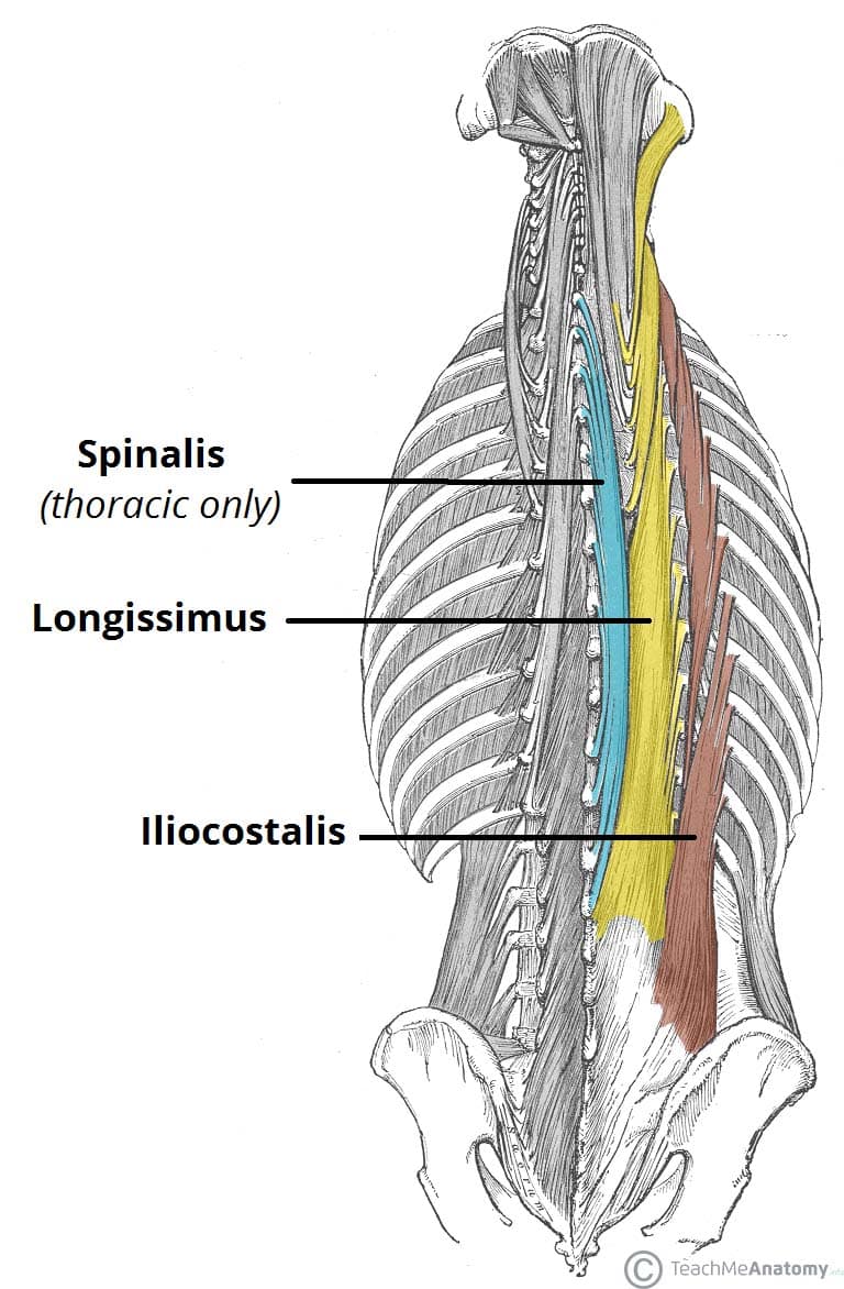
Superficial, intermediate, deep and deepest layers.these muscles lie on each side of the vertebral column, deep to the thoracolumbar fascia they span the entire length of the vertebral column, extending from the cranium to the pelvis
There are three hamstring muscles, all of them originating at the ischial tuberosity (the bones you sit on): These muscles include the large paired muscles in the lower back called erector spinae which help hold up the spine and gluteal muscles. Back muscle diagrams labeled, lower back muscle diagrams labeled, human muscles, back muscle diagrams labeled, lower back muscle diagrams labeled. It also helps in extension and lateral flexion of the lumbar spine. Large muscles and an intricate network of ligaments in your lower back support serve to stabilize your spine and power your twisting and bending movements. Certain back muscles extend to other areas, like the shoulders, upper arms, and thighs. The back comprises the spine and spinal nerves, as well as several different muscle groups. Anatomynote.com found anatomy of back muscles diagram from plenty of anatomical pictures on the internet. Some of these muscles are quite large and cover broad areas. The back anatomy includes the latissimus dorsi, trapezius, erector spinae, rhomboid, and the teres major. It is responsible for extension,adduction, and (medial) internal rotation of the shoulder joint. These structures work together to support the body, enable a range of movements, and send messages from the brain to the. See back muscles and low back pain.
Anatomy of back muscles your back consists of three distinct layers of muscles, namely the superficial layer, the intermediate layer, and the deep layer. All activity in the body is regulated by muscle mass. The superficial and intermediate muscles do not develop in the back, and are classified as extrinsic muscles. The back functions are many, such as to house and protect the spinal cord, hold the body and head upright, and adjust the movements of the upper and lower limbs. Browse 3,579 back muscle anatomy stock photos and images available, or search for pelvic bone or lymphatic system to find more great stock photos and pictures.
Muscles Of The Back Teachmeanatomy from teachmeanatomy.info We hope this picture anatomy of back muscles diagram can help you study and research. It also helps in extension and lateral flexion of the lumbar spine. See back muscles and low back pain. Muscle mass are just how we move as well as live. On this page, you'll learn about each of these muscles, their locations and functional anatomy. Labeled anatomy chart of neck and back. Learn anatomy back labeling with free interactive flashcards. Five pairs of lumbar spinal nerves labeled l1 to l5 branch off your spinal cord and exit through small holes between the vertebrae.
Back muscle diagrams labeled, lower back muscle diagrams labeled, human muscles, back muscle diagrams labeled, lower back muscle diagrams labeled.
The back anatomy includes the latissimus dorsi, trapezius, erector spinae, rhomboid, and the teres major. We hope this picture anatomy of back muscles diagram can help you study and research. Back muscles from www.corpshumain.ca back muscle diagram labeled muscle diagram new 50 unique diagram back muscles learn. The deep back muscles, also called intrinsic or true back muscles, consist of four layers of muscles: Nerves in your lower back. There are three hamstring muscles, all of them originating at the ischial tuberosity (the bones you sit on): Back dissection, cranial neve 11, dissection, latisumus dorsi, lavator. Other muscles are small and cover much less space. These muscles include the large paired muscles in the lower back called erector spinae which help hold up the spine and gluteal muscles. These muscles include the large paired muscles in the lower back called erector spinae which help hold up the spine and gluteal muscles. Large muscles and an intricate network of ligaments in your lower back support serve to stabilize your spine and power your twisting and bending movements. Labeled anatomy chart of male back muscles on white learn more about it at kenhub. Back muscle diagram diagram of female back information schematics wiring diagrams.